


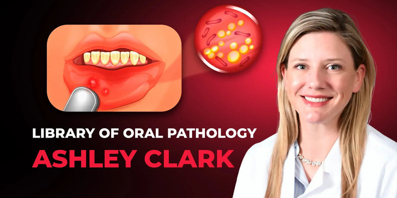
Library of Oral Pathology: Management of Precancerous and Cancerous Oral Lesions

Library of Oral Pathology: Management of Precancerous and Cancerous Oral Lesions

عن الكورس
كورس مع Dr. Ashley Clark عن تشخيص وعلاج الآفات قبل السرطانية والسرطانية بالفم. يغطي كل شي من الفحص للتشخيص النسيجي والأشعة.
مناسب لـ: طبيب الأسنان العام وأخصائي أمراض الفم
شنو راح تتعلم:
- تشخيص الـ Leukoplakia والـ Squamous Cell Carcinoma
- الـ Differential Diagnosis للتقرحات والآفات الملونة
- تشخيص وعلاج أمراض الغدد اللعابية
- قراءة الأشعة والـ Histopathology للآفات
Library of Oral Pathology: Management of Precancerous and Cancerous Oral Lesions
Course overview
Everything from diagnosis to treatment of the most common precancerous and cancerous oral diseases by Dr. Ashley Clark — A diplomate of the American Board of Oral and Maxillofacial Pathology!
During the course you will learn the protocols of:
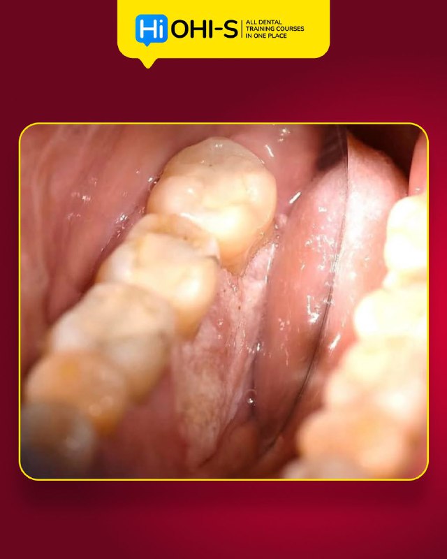
Diagnosis of leukoplakia
Detection of squamous cell carcinoma of the oral cavity
Differential diagnosis of ulcerative lesions
Diagnosis and treatment of salivary glands pathology
Diagnosis and study of histopathology
X-ray diagnosis of lesions.
The course covers all questions related to precancerous & cancerous lesions of the oral cavity and protocols for their early diagnosis and treatment.
- Differentiate clinical features of leukoplakia and other potentially malignant oral disorders.
- Formulate differential diagnoses for ulcerative and pigmented oral lesions using clinical criteria.
- Interpret radiographic features of common jaw lesions and correlate with clinical findings.
- Apply diagnostic protocols for salivary gland pathology and histopathologic assessment of oral lesions.
What you will learn
Course lessons
Lesson 1. Leukoplakia & oral cancer
Introduction to the classification and the diagnosis protocols of oral and oropharyngeal squamous cell carcinoma
The etiology of human papillomavirus
Clinical features of potentially malignant disorders of the oral cavity
Clinical features of oral dysplasia
Lesions of leukoplakia
Thin leukoplakia: hyperkeratosis, acanthosis, carcinoma-in-situ
Thick leukoplakia: mild dysplasia, mild dysplasia with hyperkeratosis
Veruciform leukoplakia: severe dysplasia
Erythroleukoplakia: severe dysplasia, squamous cell carcinoma
Common locations in which leukoplakia is encountered
The appropriate treatment plan for patients with leukoplakia
Lesions of oral squamous cell carcinoma
Methods on how to help prevent squamous cell carcinoma in your patient population.
Lesson 2. Ulcers of the oral cavity
Ulcerative conditions of the oral cavity
The differential diagnosis for ulcerative lesions
Diagnosis, treatment protocols, and case presentation of desquamative gingivitis
Differential diagnosis of desquamative gingivitis
Diagnosis, treatment protocols, and case presentation of lichen planus:
Reticular form
Papules form
Plaque-like form
Atrophic form
Ulcerative form
Bullous form.
Diagnosis, treatment protocols, and case presentation of mucous membrane pemphigoid (MMP)
Diagnosis, treatment protocols, and case presentation of Pemphigus vulgaris
The differences between acute and chronic ulcerative conditions
Diagnosis, treatment protocols, and case presentation of recurrent aphthous ulceration (RAU): minor and major form
Diagnosis, treatment protocols, and case presentation of herpes simplex virus and recurrent herpes simplex
Treatment plan for patients with acute ulcers
Treatment plan for patients with chronic ulcers
The follow-up measures for patients with oral ulcerations.
Lesson 3. Common epithelial, mesenchymal, and salivary lesions of the oral cavity
Pigmented lesions of the oral cavity and their differential diagnosis:
Amalgam tattoos: diagnosis, treatment protocols, and clinical presentation
Oral Melanotic macule: diagnosis, treatment protocols, and clinical presentation
Acquired melanocytic nevus: diagnosis, treatment protocols, and clinical presentation
Melanoma: diagnosis, treatment protocols, and clinical presentation.
Human papillomavirus: the difference between low-risk and high-risk HPV strains
The papillary lesions of the oral cavity and their differential diagnosis:
Squamous papilloma: diagnosis, treatment protocols, and clinical presentation
Verruca vulgaris: diagnosis, treatment protocols, and clinical presentation
Condyloma acuminatum: diagnosis, treatment protocols, and clinical presentation.
Differential diagnosis of mesenchymal lesions: fibromas and bumps on the gum
Salivary pathology: mucoceles and xerostomia
Treatment plan for patients with common oral lesions
Clinical cases and conclusion.
Lesson 4. Common radiographic oral pathology
Radiographic features, diagnosis, and management of radiolucent defects:
stafne defect.
Radiographic features, diagnosis, and management of multilocular radiolucent lesions:
ameloblastoma
central giant cell granuloma
hemangioma
odontogenic keratocyst.
Radiographic features, diagnosis, and management of common radiopaque lesions:
condensing osteitis
idiopathic osteosclerosis
cemento-osseous dysplasia
focal cemento-osseous dysplasia
periapical cemento-osseous dysplasia
florid cemento-osseous dysplasia
cemento blastoma
osteoma.
The difference between lesions with a similar radiographic presentation
When radiographic lesions do need a biopsy?
Follow-up measures regarding radiographic pathology
Syndromes associated with radiographic pathology.
أسئلة شائعة عن الكورس
من محاضر الكورس؟
الكورس مقدم من: Ashley Clark
كم مدة الكورس وكم درس فيه؟
الكورس يتكون من 4 درس، ومدته الكلية 3 ساعة و25 دقيقة.
هل الكورس كامل أو مجرد فيديوهات من يوتيوب؟
الكورس اشتراك كامل بكل الدروس المذكورة بالوصف (4 درس)، وليس فيديوهات يوتيوب أو مقاطع متفرقة.
متى يتفعّل الكورس بعد الدفع؟
التفعيل فوري خلال دقائق من إتمام الدفع في أغلب الحالات، وتوصلك رسالة على البريد الإلكتروني بكل تفاصيل التفعيل.
ما هي طرق الدفع المتوفرة؟
للأطباء داخل العراق: زين كاش، سوبر كي (ماستر الرافدين)، FIB أو رصيد آسيا سيل.
للأطباء خارج العراق: البطاقة البنكية، أو طرق دفع محلية في بعض الدول مثل مصر والجزائر وفلسطين وغيرها.
هل الدفع آمن؟
نعم، جميع عمليات الدفع محمية عبر بروتوكولات أمان SSL.
إذا صارت عندي مشكلة بالتفعيل، شنو الحل؟
فريق الدعم متواجد عبر واتساب أو تليغرام أو البريد الإلكتروني support@dentalcourse.store. وإذا كانت المشكلة بالتفعيل أو خلل من طرفنا، نستبدل لك الكورس.


