


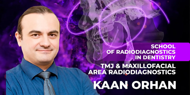
School of Radiodiagnostics in Dentistry — Module 1: TMJ & Maxillofacial Radiodiagnostics

School of Radiodiagnostics in Dentistry — Module 1: TMJ & Maxillofacial Radiodiagnostics

عن الكورس
الوحدة الأولى من مدرسة الأشعة التشخيصية بطب الأسنان مع Prof. Kaan Orhan، مخصصة للـ TMJ ومنطقة الوجه والفكين. تشرح تشريح المفصل وأمراضه وبروتوكولات التصوير وكيف تحول النتائج لخطة علاج.
مناسب لـ: أطباء الأسنان بكل الاختصاصات
شنو راح تتعلم:
- تشريح الـ TMJ الطبيعي ويا الأمراض والحالات الجهازية
- تشخيص الـ Disc Displacement والـ Degenerative Joint Disease والـ Subluxation
- بروتوكولات الـ MRI والـ CBCT والـ USG والـ Panoramic
- تحليل النتائج وإدخالها بخطة علاج اضطرابات الـ TMJ
School of Radiodiagnostics in Dentistry — Module 1: TMJ & Maxillofacial Radiodiagnostics
Course overview
This is the first module of the School of radiodiagnosis in dentistry.
From the Kaan Orhan's 10 lessons you will learn:
The anatomy of TMJ: normal, with different pathologies and systemic conditions
TMJ pathologies diagnosis: disc displacement, degenerative joint disease and subluxation
Detailed protocols of MRI, CBCT, USG, intraoral and panoramic radiography of maxillofacial area and TMJ
How to analyze received data and implement it into TMJ disorders treatment plan.
This course is recommended for dentists of all specialties.
- Identify TMJ anatomy and common pathologies across MRI, CBCT, USG, and conventional radiographs.
- Choose appropriate imaging modalities and protocols for specific TMJ and maxillofacial clinical questions.
- Interpret imaging features of disc displacement, degenerative disease, and subluxation for clinical correlation.
- Integrate radiologic findings into TMJ disorder treatment planning and communication with referring clinicians.
What you will learn
Course lessons
Lesson 1. Anatomy, histology, physiology and biomechanics of the TMJ
Radio-anatomy of the TMJ and surroundings
Normal anatomy: osseous structures and soft tissue components
Synovial membrane, capsule, ligaments
Anatomy of the disc in sagittal and coronal section
Retrodiscular lamina
TMJ vascularization
TMJ biomechanics
Clinical examination of a patient. Muscle palpation. Tender points.
Lesson 2. TMJ pathologies: disc displacement, degenerative joint disease and subluxation
Criteria of the TMJ disc(ADD) position classification. Types of disc displacements
Articular disc displacement with and without reduction
Partial articular disc displacement
Sideway disc displacement
Posterior disc displacement
Joint effusion
Bone marrow edema (osteoarthrosis)
Osteoarthritis
TMJ developmental anomalies. Hypoplasias. Bifid and trifid condyle
Traumas of the TMJ. Luxations
TMJ disorders
Erosion and cysts
Review of clinical cases.
Lesson 3. Introduction to TMJ Imaging, conventional techniques, CT/CBCT in diagnostics of TMJs
Conventional techniques in radiology
Panoramic radiography
Tomography techniques: conventional tomography, computed tomography (CT), cone beam computed tomography (CBCT)
Principles of the CBCT work. Mechanism and process. Safe doses
Specifications of the CBCT
Artifacts on the CBCT
CBCT anatomy of the maxillofacial area. Correlation with a gross anatomy
Review of clinical cases.
Lesson 4. MRI of TMJ
Timeline of MRI imagining
Fundamentals of MRI examination
Mechanism of MRI machine function. The procedure
TMJ MRI basic anatomy
TMJ disc and structure anatomy
Patterns of basic pathologies
MRI of muscles
Connections with TMJ and temporal bone and surrounding tissues.
Lesson 5. USG of TMJ
Maxillofacial ultrasound anatomy. Regions and structures
Muscles and salivary glands
Blood vessels on static and dynamic MRI
Ultrasonic scanning regarding gross anatomy
Oral floor in transverse and longitudinal sections
Normal salivary anatomy on MRI in transverse anlongitudinal sections
Normal and reactive lymph nodes on MRI
MRI and USG of the TMJ: position and structures
Indications for USG examination
Correlation of normal and reactive structures on USG and CBCT
TMJ disorders observation on USG. Correlation with MRI and CBCT images
Review of a clinical case.
Lesson 6. Intraoral radiographic technique, indications, scanning, radioanatomy
Part 1 – Theoretical
Intraoral radiography techniques
Intraoral radiography. Criteria of quality
Basic principles of projection geometry for radiography
Image sharpness and resolution. How to maximize it
Image size and shape distortion
Periapical radiography. The paralleling technique
General steps for making an exposure. Step-by-step protocol
Receptor placement during the intraoral radiography
Bisecting-angle technique. When to apply it
Donovan technique
Le Master technique
Slob technique
Vertical and horizontal bitewing receptors
Occlusal radiograph. Indications
Intraoral radiographic examination of children: mobile radiography
Special considerations.
Part 2 – Practical
Normal and pathological radioanatomy of teeth and surrounding tissues
In this part, you will learn in detail the normal and pathological radioanatomy of the teeth, periapical and surrounding tissues and will be able to analyze the radiographic images and make a diagnosis on your own.
Lesson 7. Introduction OPG: radiographic technique, indications, scanning, radioanatomy, most common pathologies
Orthopantomography. Definition and description. Historical review
Tomographic image layer
Digital system detectors in panoramic radiographs. Features
Indications for the panoramic imaging indications
Advantages and disadvantages of panoramic images
The procedure. Patient positioning. Tongue position. Mistakes during the procedure
The neck extension
Common artifacts:
Ghost image
Foreign bodies
Metal artifacts
Closing artifacts
Motion artifacts
Lead apron
Incorrect tongue position.
Anatomical landmarks: a detailed description of hard and soft tissues on the OPG
Panoramic imaging indications
Diagnostics problems requiring broad coverage of the jaws
Review of different clinical situations on the OPG:
Evaluation of trauma
Location of wisdom teeth
Extensive dental or osseous diseases
Known or suspected large lesions
Tooth development
Retained teeth or root tips (in edentulous patients)
TMJ problems which involve hard tissues (condyle)
Developmental anomalies.
Pathologies on the OPG:
Cystic lesions
Bening tumors
Malignant tumors
Fibro-osseus lesions
Oseonecrosis (osteomyelitis/MRONJ/ORN).
Considerations of different clinical cases.
Lesson 8. Introduction to CBCT radiographic technique, indications, scanning, radioanatomy, most common pathologies
Basics of CBCT
Common pathologies on CBCT
The principles of operation of dentomaxillofacial CBCT units and inherent differences in their x-ray detection systems
The artifacts
Major differences between OPG and CBCT.
Lesson 9. Introduction to MRI radiographic technique, indications, scanning, radioanatomy, most common pathologies
Basic knowledge of MRI
Application of knowledge of anatomy, physiology, positioning on MRI
Common pathologies on MRI
The artifacts
Determining imaging parameters to achieve optimum imaging, application of ferromagnetic safety principles and contrast application for the protection of the patient, technologist, and others.
Lesson 10. Introduction to USG imaging, radiographic technique, indications, scanning, radioanatomy, most common pathologies
Basic knowledge of USG
Transducion positions to scan DMFR area and surrounding tissues
Pathology recognition on USG.
أسئلة شائعة عن الكورس
من محاضر الكورس؟
الكورس مقدم من: Kaan Orhan
كم مدة الكورس وكم درس فيه؟
الكورس يتكون من 10 درس، ومدته الكلية 13 ساعة و24 دقيقة.
هل الكورس كامل أو مجرد فيديوهات من يوتيوب؟
الكورس اشتراك كامل بكل الدروس المذكورة بالوصف (10 درس)، وليس فيديوهات يوتيوب أو مقاطع متفرقة.
متى يتفعّل الكورس بعد الدفع؟
التفعيل فوري خلال دقائق من إتمام الدفع في أغلب الحالات، وتوصلك رسالة على البريد الإلكتروني بكل تفاصيل التفعيل.
ما هي طرق الدفع المتوفرة؟
للأطباء داخل العراق: زين كاش، سوبر كي (ماستر الرافدين)، FIB أو رصيد آسيا سيل.
للأطباء خارج العراق: البطاقة البنكية، أو طرق دفع محلية في بعض الدول مثل مصر والجزائر وفلسطين وغيرها.
هل الدفع آمن؟
نعم، جميع عمليات الدفع محمية عبر بروتوكولات أمان SSL.
إذا صارت عندي مشكلة بالتفعيل، شنو الحل؟
فريق الدعم متواجد عبر واتساب أو تليغرام أو البريد الإلكتروني support@dentalcourse.store. وإذا كانت المشكلة بالتفعيل أو خلل من طرفنا، نستبدل لك الكورس.


