


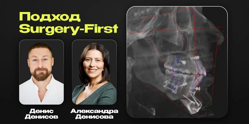
Surgery-First approach. Synchronization of orthodontics and TMJ for predictable aesthetic results

Surgery-First approach. Synchronization of orthodontics and TMJ for predictable aesthetic results

عن الكورس
كورس مع Aleksandra Denisova و Denis Denisov عن نهج الـ Surgery-First. تنسيق بين أخصائي التقويم وجراح الوجه والفكين لنتائج متوقعة.
مناسب لـ: أخصائي التقويم وجراح الوجه والفكين
شنو راح تتعلم:
- اختيار المريض ومراحل التحضير
- الـ Le Fort والـ Segmental Osteotomy
- الـ BSSO والـ Genioplasty والـ SARPE
- التأهيل بعد العملية والمتابعة التقويمية
Approach Surgery-First. Synchronizing Orthodontics and TMJ Surgery for Predictable Aesthetic Outcomes
Course overview
Surgery-First — is an interdisciplinary approach to orthognathic therapy, based on the coordinated work of the orthodontist and maxillofacial surgeon!
The course covers clinical aspects of implementing this protocol: from patient selection and preparation stages to postoperative management and orthodontic rehabilitation.
During the course you will study:
- Osteotomy protocols by Lefort and segmental osteotomy
- Protocols for bilateral sagittal osteotomy (BSSO) and genioplasty
- Protocols for surgically assisted expansion of the maxilla (SARPE)
- Protocols for postoperative rehabilitation and orthodontic follow-up
Course lessons
Lesson 1. Synchronizing orthodontists and surgeons for predictable results
- The importance of an interdisciplinary approach in modern dentistry and maxillofacial surgery
- Interdisciplinary collaboration: orthodontists, surgeons and prosthodontists
- Definition of orthodontics and orthognathic surgery
- Role of each specialist in treating patients with maxillofacial anomalies
- Types of pathologies requiring an interdisciplinary approach
- Diagnosis and treatment planning
- Types of orthognathic operations
- Preoperative preparation
- Orthodontic treatment for decompensation of the bite
Lesson 2. Le Fort osteotomy. Segmental osteotomy
Maxillary anatomy:
- Features of maxillary structure
- Blood supply and innervation
- Important anatomical landmarks
- Definition of maxillary osteotomy
- Main indications for the operation
- Indications and contraindications:
- Deformities of the craniofacial region
- Occlusal disturbances
- General and local contraindications to surgery
Diagnosis and planning of the operation:
- Clinical examination
- Radiological methods (CT, panoramic radiograph)
- Using 3D modeling for planning
Types of maxillary osteotomies:
- Le Fort I, II, III
- Segmental osteotomy
- Features of each type
Surgical technique:
- Patient preparation
- Access and steps of the operation
- Fixation of jaw fragments
- Possible complications and their prevention
Lesson 3. Bilateral sagittal osteotomy (BSSO)
- Definition of bilateral sagittal osteotomy (BSSO)
- Main indications for performing BSSO: correction of prognathism, retrognathism, open bite and other occlusal anomalies
- Mandibular anatomy: structure, blood supply, innervation
- Features of sagittal splitting: location of mandibular branches, relation to the mandibular canal
- Clinical examination: assessment of bite, facial symmetry, temporomandibular joint function
- Radiological diagnostics: orthopantomogram, computed tomography (CT), 3D modeling
- Stages of preoperative planning: fabrication of surgical templates, simulation of outcome
- Surgical technique:
- Patient preparation: anesthesia, position on the operating table
- Access to the mandible: intraoral incision, exposure of the mandibular branches
- Osteotomy steps: performing sagittal splitting, mobilization of fragments
- Fixation of fragments: use of miniplates, screws or other fixation systems
- Wound closure: suturing, drainage if necessary
Complications and their prevention:
Possible complications: damage to the inferior alveolar nerve, bleeding, infection
Prevention methods.
Postoperative period
Postoperative care: oral hygiene, diet, activity restrictions
Rehabilitation and orthodontic treatment.
Clinical cases: application of BSSO with before-and-after photos.
Lesson 4. Genioplasty in orthognathic surgery: aesthetics and function
Definition of genioplasty. Main goals and objectives of genioplasty
Anatomy and physiology of the chin region:
- Anatomical structure of the chin
- Functional role of the chin region
- Aesthetic parameters of the chin
- Aesthetic indications and functional indications for genioplasty
- Psychological aspects and patient expectations
- Types of genioplasty:
- Augmentative genioplasty
- Reduction genioplasty
- Chin asymmetry correction
- Reconstructive genioplasty
Surgical and non-surgical methods of performing genioplasty
Preoperative preparation:
- Diagnostics and planning
- Patient examination
- 3D modeling and computer planning
Technique of the operation:
- Stages of the surgical procedure
- Materials and instruments used
- Possible complications and their prevention
Postoperative period:
Possible side effects
Recommendations for patients.
Alternative methods of chin correction. Advantages and disadvantages of genioplasty.
Lesson 5. Intraoperative aspects and management of complications
- Main types of complications associated with intraoral methods
- Anatomical and physiological features of the oral cavity influencing complications
- Classification of complications in orthognathic surgery:
- Early complications (bleeding, infection, edema)
- Late complications (deformity recurrence, bite dysfunction, neurological issues)
- Intraoral access-related complications (soft tissue, teeth, nerves damage)
Intraoral aspects of complication prevention
Management of intraoral complications:
- Bleeding management: hemostasis methods, use of hemostatic materials
- Infection prevention and treatment: antibiotic therapy, local antiseptics
- Soft tissue restoration: suture materials, closure techniques
- Neurological complication handling: nerve protection, rehabilitation methods
Rehabilitation and postoperative monitoring:
Wound healing and restoration of functions control
Bite and occlusion issues correction.
Analysis of errors and their consequences.
Lesson 6. Maxillary expansion: MARPE, SARPE
Brief overview of maxillary expansion methods
Maxillary anatomy and physiology:
Palatal suture physiology and its role in jaw widening
Age-related changes of the palatal suture and their influence on treatment planning.
Indications and contraindications for maxillary expansion:
- Clinical indications for MARPE and SARPE
- Contraindications and limitations
- Diagnosis and treatment planning
MARPE (Mini-Implant Assisted Rapid Palatal Expansion):
- MARPE principle of operation
- Device design and components
- MARPE advantages: minimal invasiveness, tissue preservation, applicability in adults
- Steps of installation and activation of the appliance
- Clinical cases and outcomes
SARPE (Surgically Assisted Rapid Palatal Expansion):
- SARPE principle of operation
- Surgical steps: osteotomy, appliance installation
- SARPE advantages: efficacy in pronounced deficiencies, applicability in patients with a closed palatal suture
- Complications and their prevention
- Clinical cases and outcomes
Comparison of MARPE and SARPE:
- Comparison of effectiveness, invasiveness, indications and contraindications
- Advantages and disadvantages of each method
- Method selection depending on patient age, deficiency severity and clinical situation
Complications and their management:
Possible complications with MARPE and SARPE
Prevention and correction methods for complications.
Lower jaw expansion protocols
Rehabilitation and postoperative follow-up:
- Rehabilitation stages after MARPE and SARPE
- Patient recommendations
- Control visits and outcome assessment
Lesson 7. Surgery First: orthodontic preparation and postoperative management
- Overview of the Surgery First (SF) approach in orthodontics and maxillofacial surgery
- Main indications and contraindications for Surgery First;
- Core principles of Surgery First:
- Definition and essence of the method
- Differences from the traditional approach (orthodontics - surgery - orthodontics)
- Patient benefits
Orthodontic preparation for Surgery First:
- Diagnosis and treatment planning
- Diagnostic models, radiographic and 3D studies
- Features of preparing the dentition
- Minimal orthodontic adjustment before surgery
- Consideration of occlusal contacts and tooth positions
- Interaction between orthodontist and surgeon during planning
Surgical stage:
- Main types of surgeries within Surgery First
- Osteotomy of the maxilla and mandible
- Genioplasty
- Role of precise planning and use of computer technologies (CAD/CAM, 3D printing)
Postoperative management:
- Early postoperative period
- Monitoring patient status
- Prevention of complications
Orthodontic treatment after surgery:
- Bite correction and tooth position adjustments
- Use of elastics and other orthodontic appliances
- Duration and stages of postoperative orthodontics
Advantages and disadvantages of the Surgery First approach:
Comparison with the traditional approach
Potential risks and complications.
Clinical case analyses: examples of Surgery First application.
Lesson 8. Rehabilitation after surgery and orthodontic follow-up
The interrelation of surgery and orthodontics in comprehensive patient treatment
Main stages of rehabilitation after surgery. Early postoperative period:
- Wound care
- Pain management
- Prevention of complications (infections, edema, bleeding)
Main stages of rehabilitation after surgery. Late postoperative period:
- Restoration of functions (chewing, speech, breathing)
- Physiotherapy and therapeutic exercises
- Psychological support for the patient
Orthodontic follow-up after surgery:
- The role of orthodontics in postoperative rehabilitation
- Bite and tooth position correction after surgical intervention
- Use of orthodontic appliances (braces, aligners, retainers)
- Monitoring changes and adjusting the treatment plan
- Rehabilitation specifics for different types of surgeries
Role of a multidisciplinary approach:
Collaboration among surgeon, orthodontist, therapist and other specialists
Developing an individualized rehabilitation plan for each patient.
Prevention of complications and long-term monitoring:
- Regular check-ups and diagnostics
- Oral hygiene recommendations
- Maintaining treatment results
أسئلة شائعة عن الكورس
من محاضر الكورس؟
الكورس مقدم من: Aleksandra Denisova, Denis Denisov
كم مدة الكورس وكم درس فيه؟
الكورس يتكون من 8 درس، ومدته الكلية 9 ساعة و33 دقيقة.
هل الكورس كامل أو مجرد فيديوهات من يوتيوب؟
الكورس اشتراك كامل بكل الدروس المذكورة بالوصف (8 درس)، وليس فيديوهات يوتيوب أو مقاطع متفرقة.
متى يتفعّل الكورس بعد الدفع؟
التفعيل فوري خلال دقائق من إتمام الدفع في أغلب الحالات، وتوصلك رسالة على البريد الإلكتروني بكل تفاصيل التفعيل.
ما هي طرق الدفع المتوفرة؟
للأطباء داخل العراق: زين كاش، سوبر كي (ماستر الرافدين)، FIB أو رصيد آسيا سيل.
للأطباء خارج العراق: البطاقة البنكية، أو طرق دفع محلية في بعض الدول مثل مصر والجزائر وفلسطين وغيرها.
هل الدفع آمن؟
نعم، جميع عمليات الدفع محمية عبر بروتوكولات أمان SSL.
إذا صارت عندي مشكلة بالتفعيل، شنو الحل؟
فريق الدعم متواجد عبر واتساب أو تليغرام أو البريد الإلكتروني support@dentalcourse.store. وإذا كانت المشكلة بالتفعيل أو خلل من طرفنا، نستبدل لك الكورس.


